✓ 24/7 automatic processing of online orders
✓ Knowledgeable and professional Product & Technical Support
✓ Fast and reliable (re)-ordering
miRCURY Exosome Cell/Urine/CSF Kit
Cat. No. / ID: 76743
✓ 24/7 automatic processing of online orders
✓ Knowledgeable and professional Product & Technical Support
✓ Fast and reliable (re)-ordering
Features
- Excellent recovery of exosomes and other extracellular vesicles
- Easy and straightforward protocol that takes less than 2 hours
- No ultracentrifugation or phenol/chloroform steps required
- Fully compatible with the miRCURY LNA miRNA PCR System
- Suited for a variety of applications, such as miRNA or RNA profiling
Product Details
miRCURY Exosome Kits enable high-quality and scalable exosome isolation with an easy protocol that does not require special laboratory equipment. The miRCURY Exosome Serum/Plasma Kit is optimized for serum and plasma samples, while the miRCURY Exosome Cell/Urine/CSF Kit is designed for processing cell-conditioned media, urine and CSF samples. Both kits provide high exosomal recovery and seamless integration with different downstream assays.
Need a quote for your research project or would you like to discuss your project with our specialist team? Just contact us!
Performance
Efficient exosome isolation from serum and plasma
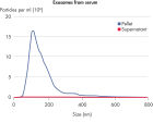
The miRCURY Exosome Serum/Plasma Kit is optimized for isolation of exosomes from serum and plasma samples. This kit includes thrombin for efficient isolation of exosomes from plasma. To evaluate the efficiency of exosome isolation, we quantified the fraction of exosomes found in the pellet versus the supernatant, which is normally discarded (see figure Exosome recovery from serum). We observed a very high enrichment rate of extracellular vesicles.
Efficient exosome isolation from cell-conditioned media, urine and CSF
The miRCURY Exosome Cell/Urine/CSF Kit is optimized for isolation of exosomes from cell-conditioned media, urine and CSF samples. In studies comparing the fraction of exosomes in the pellet versus supernatant for these sample types, we observed a very high exosome enrichment rate (see figure Exosome recovery from cell-conditioned media and Exosome recovery from urine).
Typically, the amount of miRNA present in urine and CSF samples is very limited in healthy individuals. Therefore, we highly recommend performing an exosome isolation of these biofluids prior to RNA isolation and PCR profiling. This allows you to use the exosome isolation kit as a way of concentrating the sample, while keeping the amount of potential PCR inhibitors to a minimum (see figure Detect more miRNAs in biofluids using exosome isolation kits).
See figures
Principle
Although they are not species specific, the kit protocols are optimized for working with human biofluids. Refer to the miRCURY Exosome Kits Handbook for recommended starting material for different sample types.
Why study exosomes?
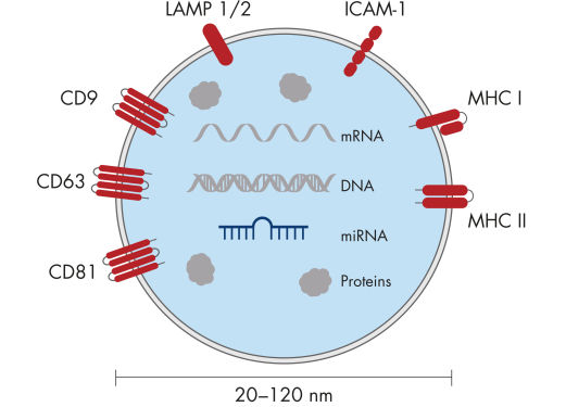
Exosomes are cell-derived, membranous particles ranging in size from 20–120 nm, approximately the same size as viruses but considerably smaller than microvesicles (see figure The structure of an exosome). Exosomes are excreted from cells into the surrounding media and can be found in many, if not all, body fluids. Their proposed role as intercellular hormone-like messengers, together with their stability as carrier of proteins and RNA, make them ideal as biomarkers for a variety of diseases and biological processes (see figure The hypothesis of exosomal shuttling of miRNAs).Exosomes are secreted by most cell types and are formed by the fusion of multivesicular bodies with the plasma membrane. They are believed to be involved in a number of functions, including:
- Immune regulation (e.g., tumor-derived exosomes may help tumors evade the immune response)
- Blood coagulation
- Cell migration
- Cell differentiation
- Cell-to-cell communication
Microvesicles that are larger than exosomes (up to 1 µm) are typically formed by blebbing of the plasma membrane, whereas exosomes are released via exocytosis from multivesicular bodies of the endosome.
See figures
Procedure
Applications
Supporting data and figures
The structure of an exosome.


